Elevated Poster Presentation International Celiac Disease Symposium 2026
PREDICTORS OF PROGRESSION TO VILLOUS ATROPHY IN CHILDREN WITH TYPE 1 DIABETES AND POTENTIAL CELIAC DISEASE (#73)
Background: Children with Type1diabetes (T1DM) and positive celiac serology but normal duodenal mucosa (potential celiac disease- CeD) often have spontaneous normalisation of serology despite a gluten-containing diet. This results in either repeated biopsies or an upfront advice for gluten-free diet (GFD) if TTG is >10ULN.
Objectives: To find the predictors of villous atrophy in children with T1DM and potential CeD.
Design: Ambispective observational; Children with T1DM and potential CeD on a gluten-containing diet were included. Clinical, serological, histological, and immunohistochemical parameters at baseline and follow-up biopsy were evaluated.
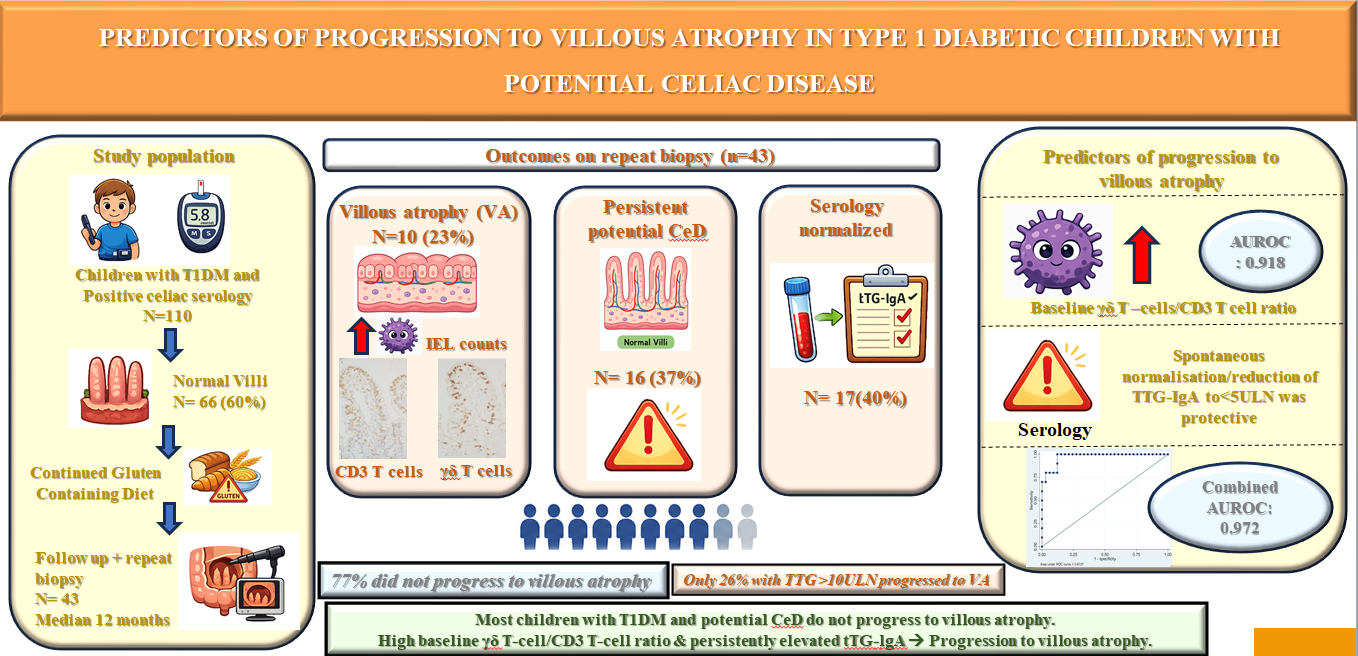
Results: Of the 110 children with T1DM and positive celiac serology, 66(60%) were potential CeD. Forty-three children had follow-up and repeat biopsy after a median of 12 months; 10 (23%) developed villous atrophy(VA), 16(37%) remained potential CeD, and 17(40%) had spontaneous normalisation of serology. Children who developed VA had higher baseline counts of intraepithelial lymphocytes(IELs), γδ T cells, and CD3 T cells. In multivariate analysis, the baseline γδ T cell/CD3 T cell ratio was an independent predictor of progression to VA(OR 1.15; 95% CI 1.03–1.28; p=0.011; AUROC 0.918). Spontaneous normalisation/reduction of TTG-IgA to <5ULN was associated with reduced risk of VA(OR 0.007, 95% CI 0.00007–0.617; p = 0.030); Combined AUROC for both was 0.972. VA developed in only 26%(6/23) with TTG >10 ULN at baseline.
Conclusions: Most children with T1DM & positive celiac serology do not progress to VA; Initiating a GFD without biopsy solely on the basis of TTG>10ULN may be unjustified. Baseline elevation in γδ T–cells/CD3 T cell ratio and persistently high serological titres help identify the risk of progression to VA.