Oral Presentation International Celiac Disease Symposium 2026
Digital and Optical Image Analysis of Gluten-Dependent Small-Intestinal Celiac Biopsies: Bridging Quantitative and Qualitative readouts (#88)
Background and aims: Qualitative Marsh-Oberhuber (M-O) classification remains the reference standard for evaluating celiac disease (CeD) duodenal biopsies, but interobserver variability is high, prompting increased use of quantitative morphometry in research and drug trials. We aimed to (i) determine the reliability of continuous villus-to-crypt ratio (VH:CrD) morphometry and derive M-O cut-offs anchored to non-CeD reference values; (ii) establish concordance between light- and digital-microscopy readings; and (iii) determine whether M-O 2-3c biopsies consistently show concurrent inflammation.
Methods: H&E-stained slides spanning flat to normal mucosa were blindly re-evaluated by two experienced readers using prior light-microscopy VH:CrD results. CD3+ intraepithelial lymphocyte (IEL) density measured inflammation. Whole-slide scans were used for calibrated digital measurement of VH and CrD in all readable units, following a standardized operating procedure. A global VH:CrD result required at least three measurable units. In total, 144 duodenal biopsies from 54 subjects were selected, including unreadable samples. Intra- and interobserver variation was analyzed by Bland-Altman and intraclass correlation (ICC); 77 non-CeD samples from 37 subjects served as controls.
Results: Both readers agreed on 49 unmeasurable samples, mostly lacking longitudinally cut crypts. Intraobserver VH:CrD mean difference was −0.072 (ICC 0.967); interobserver difference was −0.040 (ICC 0.943). Non-CeD controls averaged VH:CrD 2.8 (95% CI 2.0–3.6). Optical-digital concordance was strong for VH:CrD (ICC 0.983) and IEL density (0.970). Among M-O 2–3c samples, 28% showed normal IEL density (<25/100 epithelial cells). Conversion of the morphometry results to qualitative M-O classes is shown in Table.
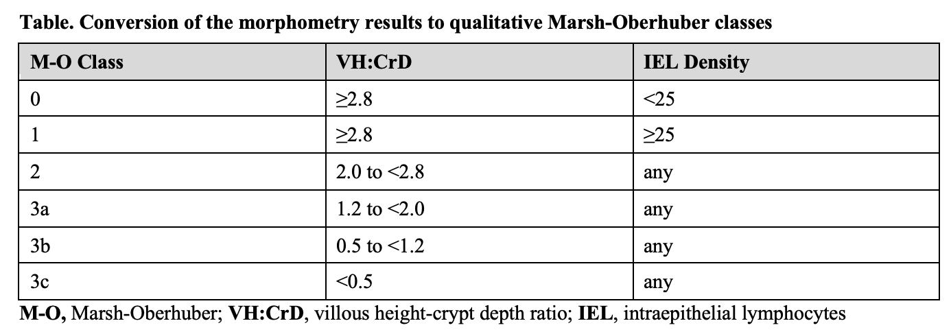
Conclusions: Substantial VH:CrD variation occurs within a single M-O class, and architectural damage and inflammation diverge in 28% of M-O 2–3c biopsies, both invisible to categorical grading. Digital morphometry matched optical reading in reliability and reproducibility, supporting centralized, standardized morphometric measurement in celiac drug trials.
- Al-Toma A, Zingone F, Branchi F, et al. European Society for the Study of Coeliac Disease 2025 Updated Guidelines on the Diagnosis and Management of Coeliac Disease in Adults. Part 1: Diagnostic Approach. United European Gastroenterol J. 2025;13(10):1855-1886.
- Werkstetter KJ, Korponay-Szabó IR, Popp A, et al. Accuracy in Diagnosis of Celiac Disease Without Biopsies in Clinical Practice. Gastroenterology. 2017;153(4):924-935.
- Taavela J, Koskinen O, Huhtala H, et al. Validation of morphometric analyses of small-intestinal biopsy readouts in celiac disease. PLoS One. 2013;8(10):e76163.